
 Part of Tibidabo Scientific Industries
 Advancing Optical BioImaging
Advancing Optical BioImaging: Bio, Chemi, and Fluorescence Technologies Combined with Irradiation Equipment.
Bioluminescence, chemiluminescence, and fluorescence are fascinating phenomena that involvethe emission of light through biological processes, chemical reactions, or in response to excitationlight, respectively (Figure 1).
Bioluminescence is the production and emission of light by living organisms, resulting frombiochemical reactions between luciferins (light-emitting molecules) and luciferases (enzymes thatcatalyse the reaction). Chemiluminescence refers to light emission caused by a chemical reaction,occurring in both biological and non-biological contexts.
Fluorescence imaging involves the emission of light from excitable fluorophores, which absorb lightat a shorter wavelength and re-emit it at a longer wavelength. This results in characteristicfluorescence, enabling the creation of detailed images of biological structures and processes.
Each method offers unique advantages, and their combination covers nearly the full range of opticalimaging, providing powerful tools for studying molecular imaging in biological phenomena.Combining these with preclinical therapeutic irradiation opens up an exceptionally wide range ofnovel scientific experiments.
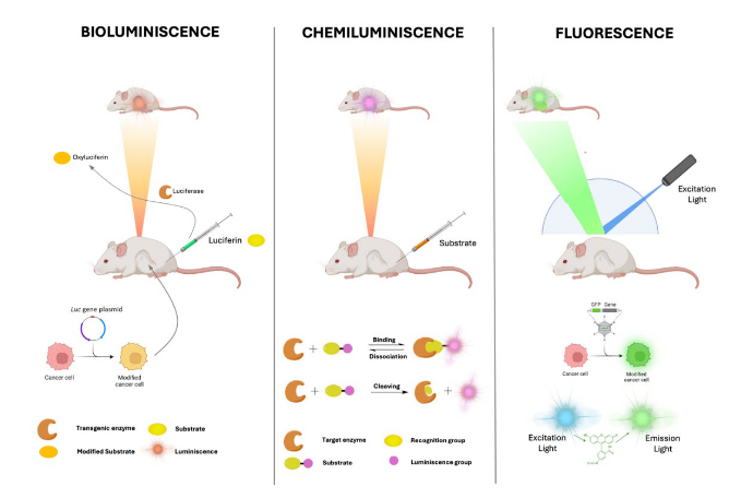
Figure 1. Overview of bioluminescence (left), chemiluminescence (middle) and Fluorescence (right) processes.Created with BioRender.com
Optical imaging capabilities in Precision X-Ray, Inc. systems: SmART+ (Small AnimalRadiotherapy System) and X-Rad Irradiators.
The Precision X-Ray, Inc. SmART+ (small animal image-guided radiotherapy) system and X-Radfamily of cabinet biological irradiators are state-of-the-art preclinical irradiators, which can beenhanced with advanced imaging capabilities through optional molecular imaging modules(OptiMAX and OptiFLEX), offering optimized tumor detection, an enhanced imaging workflow, andautomated co-registration with anatomical imaging. The integration of Optical Imaging supports awide range of applications, for example: Preclinical Imaging of Tumor Response to Radiation,Evaluating the Efficacy of Novel Drug Treatments, and Tumor Microenvironment and TherapeuticResponses 1-3 .
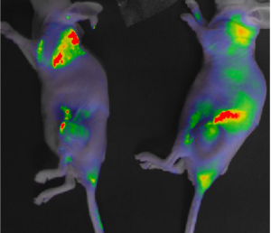
Figure 2. (left) Examples of imaging‐targeting in the SmART+ system using CT and BLI. The overlay of cellular activity measured by BLI on the CT scan is shown to optimize irradiation at the most active site of the subcutaneous (sc) tumors. (Right) FLI imaging with the prototype of the Integrated System in Precision X‐Ray equipment, showing the fusion of white light and fluorescence images.
Conclusion
The integration of these technologies (Bio, Chemi, and Fluorescence) covering the entire spectrumof optical imaging with the SmART+ and X-Rad systems (enhanced by OptiMAX and OptiFLEX), marksa significant improvement in the imaging capabilities of these systems. This integration facilitatestargeted radiation delivery and allows for effective monitoring of processes and their efficiency.Additionally, these imaging capabilities can be employed independently of radiation for researchpurposes. This combination enhances sensitivity, resolution, and workflow efficiency, facilitating abroad spectrum of applications in preclinical research.
References
1. Ji, J., Ding, K., Cheng, B., Zhang, X., Luo, T., Huang, B., … & Chen, G. (2024). Radiotherapy‐Induced AstrocyteSenescence Promotes an Immunosuppressive Microenvironment in Glioblastoma to Facilitate TumorRegrowth. Advanced Science, 11(15), 2304609.
2. Douyère, M., Gong, C., Richard, M., Pellegrini‐Moïse, N., Daouk, J., Pierson, J., … & Boura, C. (2022). NRP1inhibition modulates radiosensitivity of medulloblastoma by targeting cancer stem cells. Cancer CellInternational, 22(1), 377.
3. Daouk, J., Jubréaux, J., Chateau, A., Schohn, H., & Pinel, S. (2020). Imaging performance of a multimodalmodule to enhance preclinical irradiator capabilities. Clinical Oncology and Research, 3(2)
