
 Part of Tibidabo Scientific Industries
 Basics of Cone Beam Computed Tomography
Basics of Cone Beam Computed Tomography (CBCT)
Cone Beam Computed Tomography (or Cone Beam CT/CBCT) is an imaging technique to view 3dimensional images of a patient or specimen’s internal anatomy. It functions by reconstructing alarge number of 2 dimensional images, acquired using an x-ray source and a flat panel detector,into a single 3D image. These 2D images, also called projections, are acquired sequentially as thegantry moves around the specimen, which is placed along the gantry’s axis of rotation.Simultaneously, they are reconstructed into a single 3D image using an algorithm called filtered backprojection.
Clinical applications of CBCT include:
- Diagnostic imaging
- Image-guided radiation therapy
- Image-guided surgery
The SmART+ –Small Animal Radio Therapy System (Precision, X-Ray, Inc., Madison, CT) employs CBCT for the second application – Image-Guided Radiation Therapy. The 3-D images of the animal are used to precisely place the radiation target where you want to treat the animal(typically the tumor, though not always). This enables you to more precisely irradiate the targeted structures, while avoiding healthy tissues and organs at risk. Images can also be imported into a Treatment Planning System, which provides an even more precise method of delivering radiation therapy by overlaying the dose calculations on top of the images.
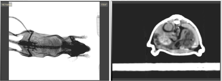
Fig 1. (left) 2D images of a plastinated mouse being acquired on the SmART+ system. (right) the respective reconstructed 3D CBCT image.
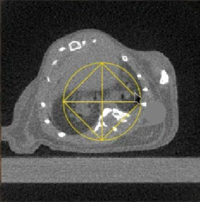
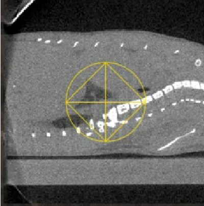
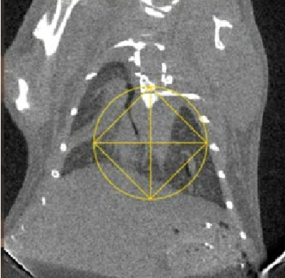
Fig 2. Reconstructed CBCT image of a mouse with lung fibrosis as viewed in the SmART+ software. (Left) Axial, (Center) Sagittal and (Right) Coronal views.
References:
1. Feldkamp, L. A., Davis, L. C. & Kress, J. W. Practical cone-beam algorithm. J. Opt. Soc. Am. A,JOSAA 1, 612–619 (1984).
2. Jaffray, D. A., Siewerdsen, J. H., Wong, J. W. & Martinez, A. A. Flat-panel cone-beam computedtomography for image-guided radiation therapy. International Journal of RadiationOncology*Biology*Physics 53, 1337–1349 (2002).
3. Clarkson, R. et al. Characterization of image quality and image-guidance performance of apreclinical microirradiator. Med Phys 38, 845–856 (2011).
