X-RAD OPTIONS
Learn more ›OptiMAX
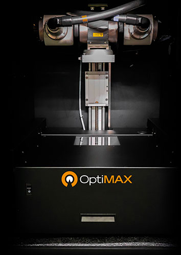
A 2D image-guided radiotherapy module that can be integrated into an X-Rad system, enabling researchers to perform multi-modality imaging with fluorescence and bioluminescence.
key Features
- Image animals in both x-ray mode and optical mode
- Combines images for co-registration of luminescent tumors or target tissue on an x-ray background
- Used in pre-treatment image-guided localization of target tissues
- Used in post-treatment diagnostics at a molecular and functional level
- Provides precise localization and documentation of high-energy collimated dose fields
- Compatible with x-ray luminescent nanoparticle imaging
 Download Brochure
 Powered by Bioz
Applications
Longitudinal Molecular Imaging
Radioisotopic Imaging
Multi-Animal Imaging and Treatment Targeting
Tumor Progression
Treatment Alignment Verification
Tumor Volume Calculation
The research which is now possible with this machine, the X-Rad320, has the potential to change cancer treatment regimens, reduce side effects, and improve quality of life.
Professor Pam Sykes
Flinders Centre for Innovation in Cancer